Contents
- 1 Unveiling the Intricacies of Our Visual System
- 2 Prof. Aécio D’Silva, Ph.D AquaUniversity
- 3 The Anatomy of the Human Eye
- 4 The Cornea and Lens
- 5
- 6 The Retina and Photoreceptors
- 7 The Optic Nerve
- 8 The Formation and Alignment of Cells
- 9 Genetic Instructions
- 10
- 11 Signaling Pathways
- 12 The Consequences of Misalignment
- 13 Retinal Detachment
- 14 Macular Degeneration
- 15
- 16 Understanding the Retina and Maintaining Good Eye Health
- 17
- 18 Maintaining Good Eye Health
- 19 Conclusion
- 20 References
Unveiling the Intricacies of Our Visual System
Prof. Aécio D’Silva, Ph.D
AquaUniversity
Summary: The human eye is a complex organ composed of trillions of cells that work together to capture and interpret visual signals. This blog explores the formation and alignment of these cells, the structure of the eye, and the consequences of misalignment, providing a comprehensive understanding of how our eyes achieve such remarkable precision.
Human eye – The human eye, a marvel of biological engineering, consists of trillions of cells perfectly aligned to capture and interpret visual signals. This intricate structure forms rapidly during development, achieving a level of precision that allows us to see the world in all its glory.
The human eye is often described as one of the most complex organs in the body. Although some try unsuccessfully to explain its evolution, analyzing its complexity it is easy to reach the scientific conclusion that it is impossible that the human eye could have evolved. It‘s so precise and orderly that it had to be created by a wonderful clever design that knows exactly what comes before and after! Its parts are so connected and dependent on each other that one part can’t evolve in isolation.
The Anatomy of the Human Eye
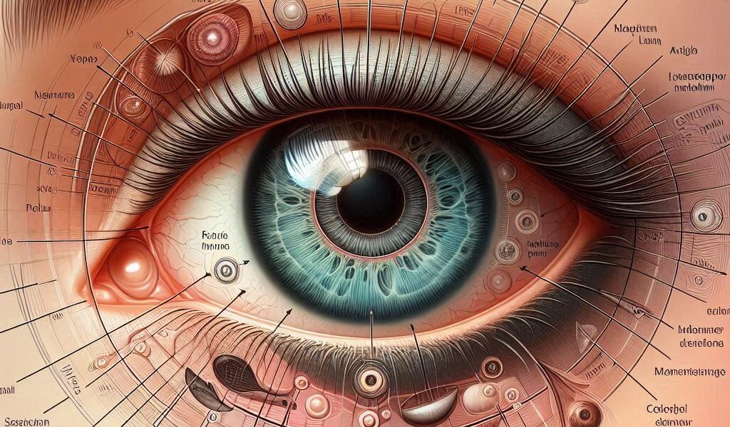
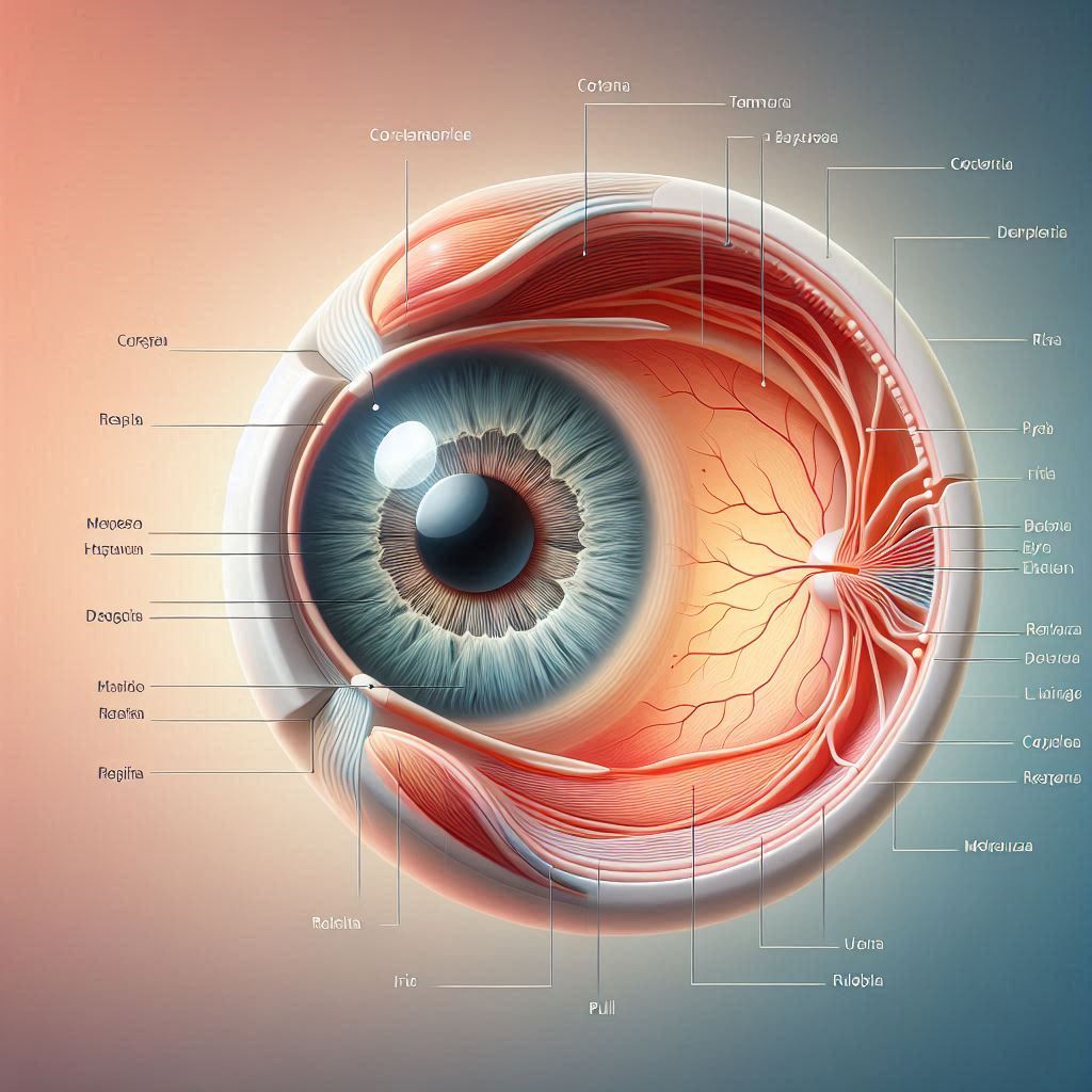
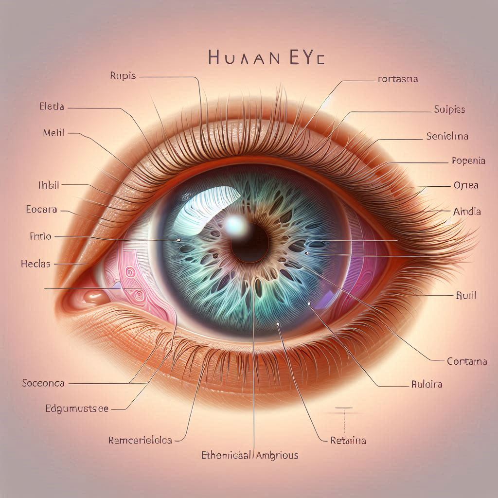
The human eye is composed of several key structures, each playing a vital role in the process of vision. These structures include the cornea, lens, retina, and optic nerve. The cornea and lens focus light onto the retina, where photoreceptor cells convert light into electrical signals. These signals are then transmitted to the brain via the optic nerve, allowing us to perceive images. The possibility of all this complexity happening by chance or evolution is practically zero.
The Cornea and Lens
The cornea is the transparent, dome-shaped surface that covers the front of the eye. It helps to focus incoming light onto the lens, further refining this focus to project a clear image onto the retina. The lens is flexible, allowing it to change shape and adjust focus for objects at different distances.
The Retina and Photoreceptors
The retina is a thin layer of tissue at the back of the eye that contains millions of photoreceptor cells, known as rods and cones. Rods are responsible for vision in low light conditions, while cones detect color and detail in brighter light. These photoreceptors convert light into electrical signals that are sent to the brain.
The Optic Nerve
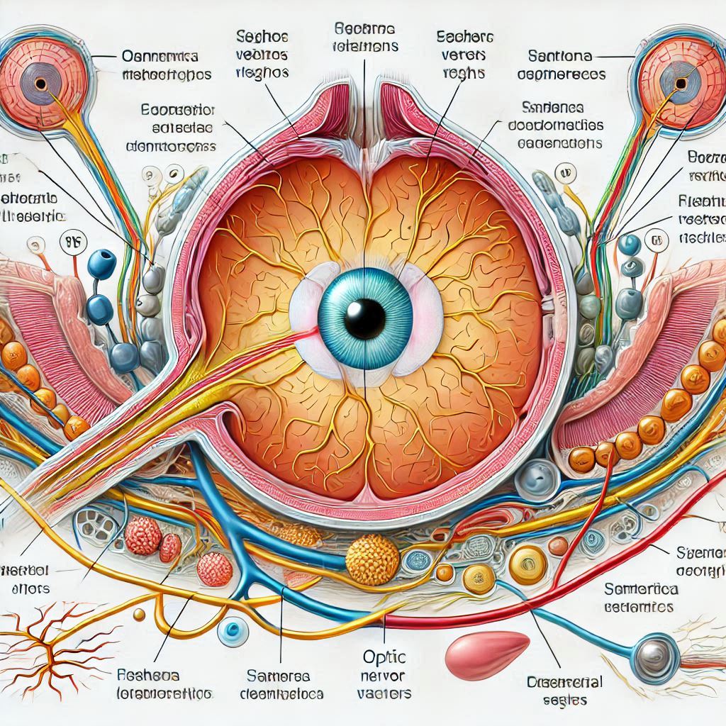
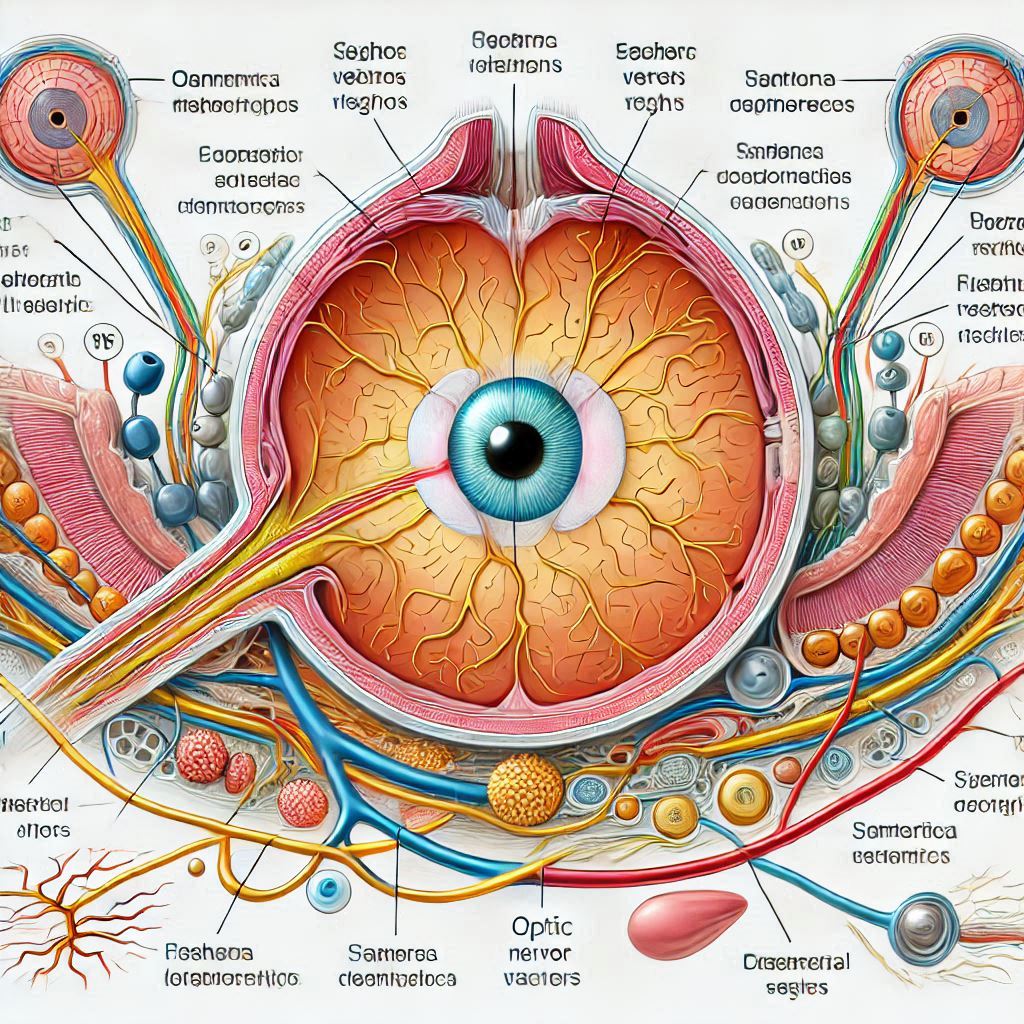
The optic nerve is a bundle of more than a million nerve fibers that transmit visual information from the retina to the brain. This transmission is crucial for the interpretation of visual signals, allowing us to understand and interact with our environment.
The Formation and Alignment of Cells
The formation and alignment of cells in the eye begin early in embryonic development. Stem cells differentiate into various types of cells, including photoreceptors, neurons, and supporting cells. This process is guided by genetic instructions and signaling pathways that ensure each cell type forms in the correct location and orientation.
Genetic Instructions
Genes play a crucial role in the development of the eye. They provide the blueprint for the formation of different cell types and their precise arrangement. Mutations in these genes, as were sustained by evolution, can lead to developmental disorders and vision problems.
Signaling Pathways
Signaling pathways are networks of molecules that communicate instructions for cell growth, differentiation, and alignment. These pathways ensure that cells form in the correct sequence and position, contributing to the overall structure and function of the eye.
The Consequences of Misalignment
Even a slight misalignment of cells in the eye can have significant consequences. Misaligned cells can disrupt the transmission of visual signals, leading to vision impairments or blindness. Conditions such as retinal detachment, macular degeneration, and congenital blindness are often the result of such misalignments.
Retinal Detachment
Retinal detachment occurs when the retina separates from the underlying tissue, disrupting the alignment of photoreceptors and leading to vision loss. This condition requires immediate medical attention to prevent permanent blindness.
Macular Degeneration
Macular degeneration is a condition where the central part of the retina, known as the macula, deteriorates. This leads to a loss of central vision and can severely impact daily activities such as reading and driving.
Understanding the Retina and Maintaining Good Eye Health
The retina is a crucial part of the eye, responsible for converting light into neural signals that the brain interprets as visual images. Let’s dive deeper into its function and explore some tips for maintaining good eye health.
The Function of the Retina
The retina is a thin layer of tissue located at the back of the eye. It contains millions of photoreceptor cells which are specialized neurons. Their primary role is to convert light into electrical signals that can be interpreted by the brain, enabling vision. There are two main types of photoreceptor cells: rods and cones.
- Rods: These cells are highly sensitive to low light levels and are crucial for night vision. They do not detect color but are excellent at sensing movement and providing peripheral vision.
- Cones: These cells function best in bright light and are responsible for detecting color. There are three types of cones, each sensitive to different wavelengths of light (red, green, and blue), which together enable color vision.
Photoreceptors play a vital role in the visual process by capturing light and initiating the complex process of visual phototransduction, where light is converted into neural signals
When light enters the eye, it is focused by the cornea and lens onto the retina. The photoreceptors then convert this light into electrical signals, which are transmitted to the brain via the optic nerve. The brain processes these signals to create the images we see.
How do photoreceptors connect to the optic nerve?
Photoreceptors in the retina connect to the optic nerve through a series of intermediate neurons. Here’s a simplified overview of the process:
- Photoreceptors (Rods and Cones): These cells detect light and convert it into electrical signals.
- Bipolar Cells: The electrical signals from the photoreceptors are transmitted to bipolar cells, which act as intermediaries.
- Ganglion Cells: Bipolar cells then pass the signals to ganglion cells. The axons of these ganglion cells converge to form the optic nerve.
- Optic Nerve: The optic nerve carries the visual information from the retina to the brain, where it is processed and interpreted.
This pathway ensures that the light detected by the photoreceptors is efficiently transmitted to the brain, allowing us to see.
How does the brain interpret visual signals from the optic nerve?
The brain interprets visual signals from the optic nerve through a complex and fascinating process involving several key structures:
- Optic Nerve: The journey begins when the optic nerve carries electrical signals from the retina to the brain. These signals are the result of light being converted into neural impulses by photoreceptor cells.
- Optic Chiasm: At the optic chiasm, some of the nerve fibers cross over to the opposite side of the brain. This crossing allows for binocular vision, which is essential for depth perception.
- Optic Tracts: After the optic chiasm, the signals continue along the optic tracts to the lateral geniculate nucleus (LGN) of the thalamus.
- Lateral Geniculate Nucleus (LGN): The LGN acts as a relay station, processing and organizing the visual information before sending it to the visual cortex.
- Visual Cortex: Located in the occipital lobe at the back of the brain, the visual cortex is where the final interpretation occurs. Here, the brain processes the signals to form images, recognize patterns, and interpret colors and movements
This intricate pathway ensures that the visual information captured by the eyes is accurately interpreted, allowing us to perceive and interact with the world around us.
How does the brain interpret visual signals from the optic nerve?
The brain interprets visual signals from the optic nerve through a complex and fascinating process involving several key structures:
- Optic Nerve: The journey begins when the optic nerve carries electrical signals from the retina to the brain. These signals are the result of light being converted into neural impulses by photoreceptor cells.
- Optic Chiasm: At the optic chiasm, some of the nerve fibers cross over to the opposite side of the brain. This crossing allows for binocular vision, which is essential for depth perception.
- Optic Tracts: After the optic chiasm, the signals continue along the optic tracts to the lateral geniculate nucleus (LGN) of the thalamus.
- Lateral Geniculate Nucleus (LGN): The LGN acts as a relay station, processing and organizing the visual information before sending it to the visual cortex.
- Visual Cortex: Located in the occipital lobe at the back of the brain, the visual cortex is where the final interpretation occurs. Here, the brain processes the signals to form images, recognize patterns, and interpret colors and movements.
This intricate pathway ensures that the visual information captured by the eyes is accurately interpreted, allowing us to perceive and interact with the world around us.
Maintaining Good Eye Health
Maintaining good eye health is essential for preserving vision and preventing eye diseases. Here are some tips to help you keep your eyes healthy:
- Eat a Balanced Diet: Consuming foods rich in vitamins A, C, and E, as well as omega-3 fatty acids, can support eye health. Include leafy greens, fish, nuts, and colorful fruits and vegetables in your diet.
- Protect Your Eyes from UV Light: Wear sunglasses that block at least 99% of UVA and UVB radiation when you are outdoors. This helps prevent damage from the sun’s harmful rays.
- Reduce Screen Time: Prolonged exposure to screens can strain your eyes. Follow the 20-20-20 rule: every 20 minutes, look at something 20 feet away for at least 20 seconds.
- Get Regular Eye Exams: Regular check-ups with an eye care professional can help detect and treat eye conditions early. It’s recommended to have a comprehensive dilated eye exam every 1-2 years.
- Practice Good Hygiene: Wash your hands before touching your eyes or handling contact lenses to prevent infections. Clean your contact lenses properly and replace them as recommended.
- Stay Active: Regular exercise improves blood circulation, which can benefit your eyes by increasing the flow of oxygen and nutrients.
By following these tips, you can help maintain your eye health and enjoy clear vision for years to come.
Conclusion
The human eye is a testament of a remarkable Intelligent Design due to the complexity and precision of biological systems. The formation and alignment of trillions of cells in such a short period are nothing short of God’s miraculous. Understanding the intricate structure of the eye and the processes that ensure its proper function highlights the marvel of human vision and the importance of maintaining eye health. If you want to learn more about Intelligent Design, the books of Prof. Marcos Eberlin are remarkable for their insightful analysis, depth as well clarity and I highly recommend them.
References
- Marcos Eberlin( (2019). Foresight: How the Chemistry of Life Reveals Planning and Purpose
- Henry M. Morris, Ph.D. 2003. The Mathematical ImpossibilityOf Evolution. Acts & Facts. 32 (11).
- James Coppedge (1973). Evolution: Possible or Impossible(Zondervan, 276 pp.)
- JL Harper (1968 ) Mathematical Challenges to the Neo-Darwinian Interpretation of Evolution. A symposium, Philadelphia, April 1966. Paul S. Moorhead and Martin M. Kaplan.
- Michael J Behe(2020) A Mousetrap for Darwin: Michael J. Behe Answers His Critics
- Michael J. Behe (2005) Darwins Black Box : Biochemical Challenge to Evolution..
- Michael J. Behe. Darwin Devolves: The New Science About DNA That Challenges Evolution
- Purves, D., Augustine, G. J., & Fitzpatrick, D. (2001). Neuroscience. Sinauer Associates.
- Heckenlively, J. R., & Arden, G. B. (2006). Principles and Practice of Clinical Electrophysiology of Vision. MIT Press.
- https://eyesurgeryguide.org/understanding-the-optic-nerve-connecting-the-eye-to-the-brain/
Disclaimer: This blog post is for informational purposes only and should not be considered medical advice. Always consult with a qualified healthcare professional for any health concerns or before making any decisions related to your health.
